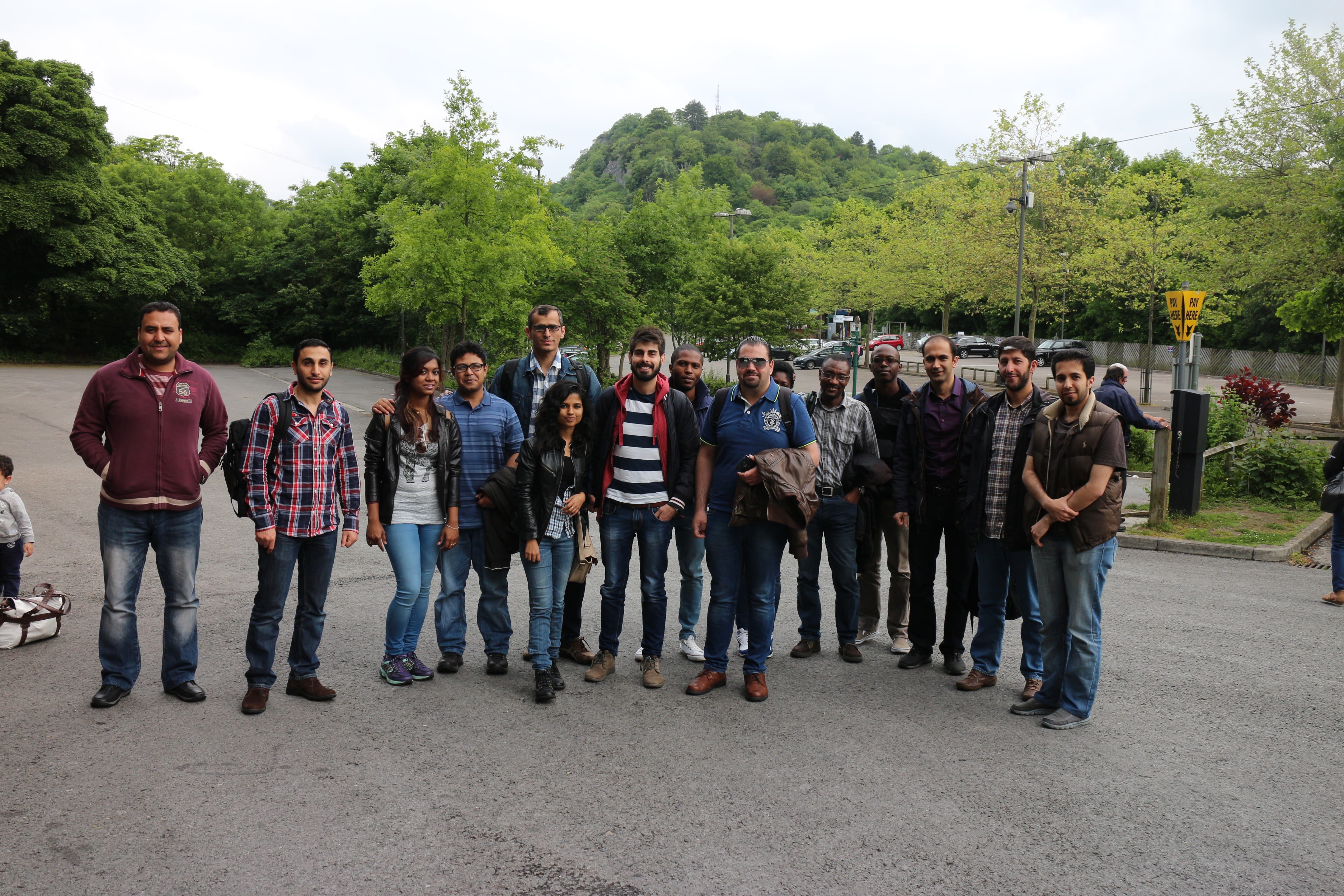
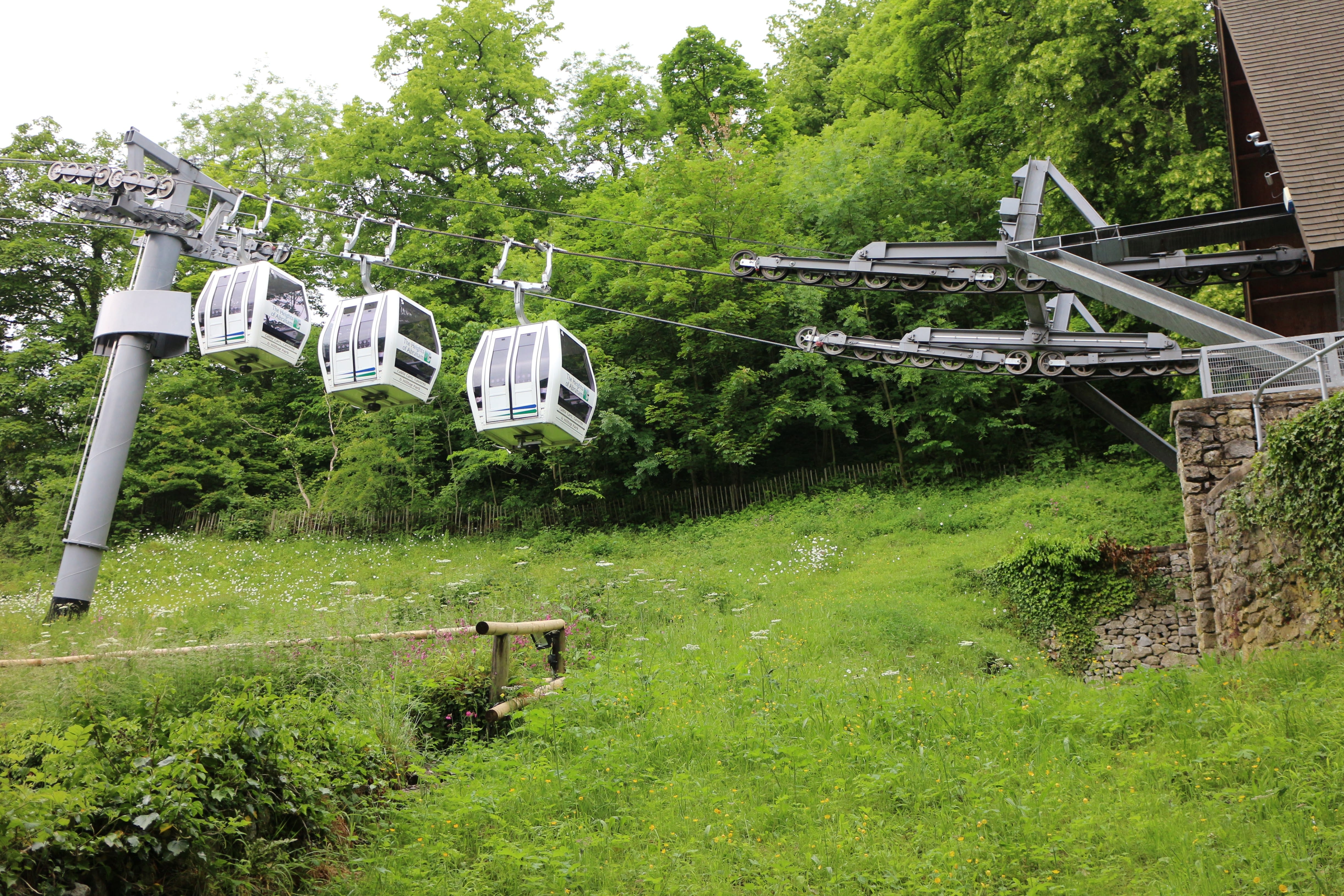
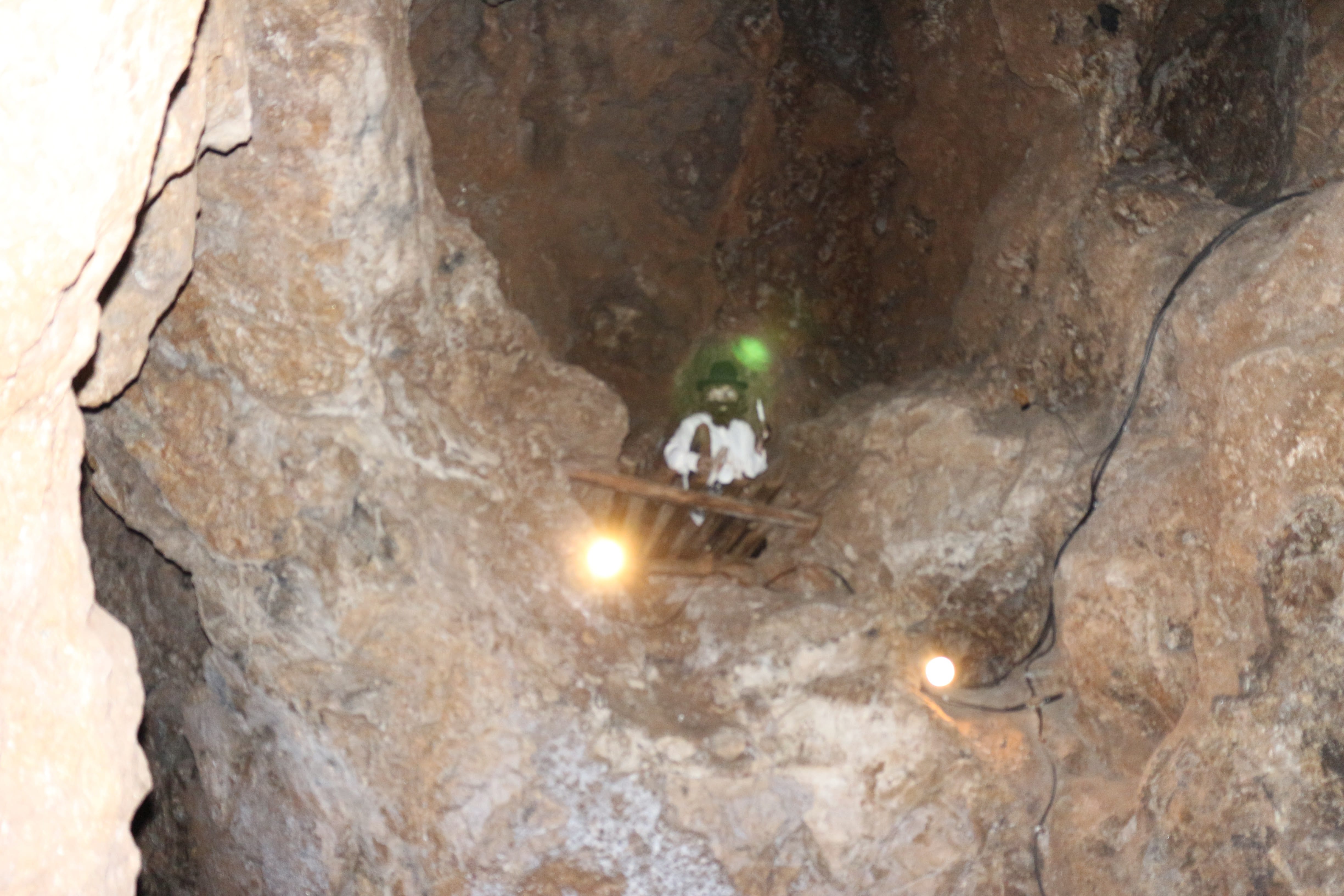
Another interesting day trip to Abraham Heights, Derby. 14th June 2015.
All got the cable cars, enjoyed tours inside old mining sites, and fantastic views from the top.
School of computer Science,
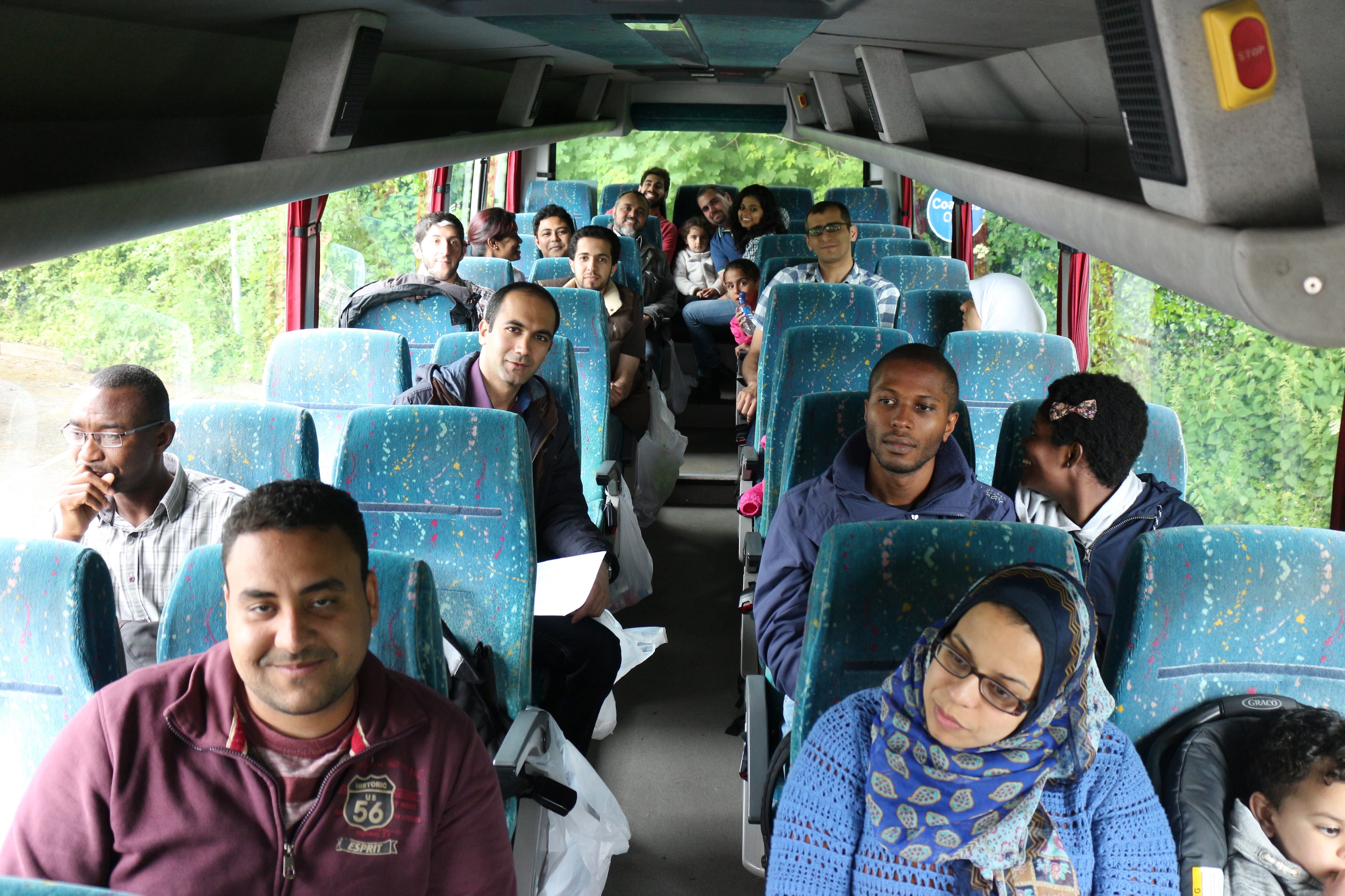
Another interesting day trip to Abraham Heights, Derby. 14th June 2015.
All got the cable cars, enjoyed tours inside old mining sites, and fantastic views from the top.
The monthly PGRs Research Presentations was held on Wed. 10th June, 2pm, Room MC3108.
This session we had the following presentations:
| Title: “Effects of Environmental Changes on Aggregation with Robot Swarm“. | Title: “Computer-aided Liver lesion detection and classification” | |
|
By: Farshad Arvin |
By: Hussein Alahmer |
|
| Abstract: Aggregation is one of the most fundamental behaviors that has been studied in swarm robotic researches for more than two decades. The studies in biology revealed that environment is a very important factor especially in cue-based aggregation in which a heterogeneity in the environment such as a heat or light source act as a cue indicating an optimal aggregation zone. In swarm robotics, studies on cue-based aggregation mainly focused on different methods of aggregation and different parameters such as population size. Although of utmost importance, effects of environmental factors have not been studied extensively. In this work, we study the effect of different environmental factors such as size and texture of aggregation cues and speed of the changes on a dynamic environment using real robots. We used aggregation time and size of the aggregate as the two metrics and evaluated the performance of the swarm aggregation in static and dynamic environments. The results of the performed experiments illustrate how environmental changes influence the performance of a swarm aggregation. |
Abstract: Liver cancer is one of the major death factors in the world. Transplantation and tumor resection are two main therapies in common clinical practice. Both tasks need image assisted planning and quantitative evaluations. An efficient and effective automatic liver segmentation is required for corresponding quantitative evaluations. Computed Tomography (CT) is highly accurate for liver cancer diagnosis. Manual identification of hepatic lesions done by trained physicians is a time-consuming task and can be subjective depending on the skill, expertise and experience of the physician. Computer aided classification of liver tumors from abdominal Computer Tomography (CT) images requires segmentation and analysis of tumor. Automatic segmentation of tumor from CT images is difficult, due to the size, shape, position and presence of other objects with the same intensity present in the image. The proposed system automatically segment liver from abdominal CT and detect hepatic lesions, then classifies the lesion into Benign or Malignant. The method uses Fuzzy C Means (FCM) clustering and region growing technique. The effectiveness of the algorithm is evaluated by comparing automatic segmentation results to the manual segmentation results. Quantitative comparison shows a close correlation between the automatic and manual as well as high spatial overlap between the regions-of-interest (ROIs) generated by expert radiologist and proposed system. |
|